product description
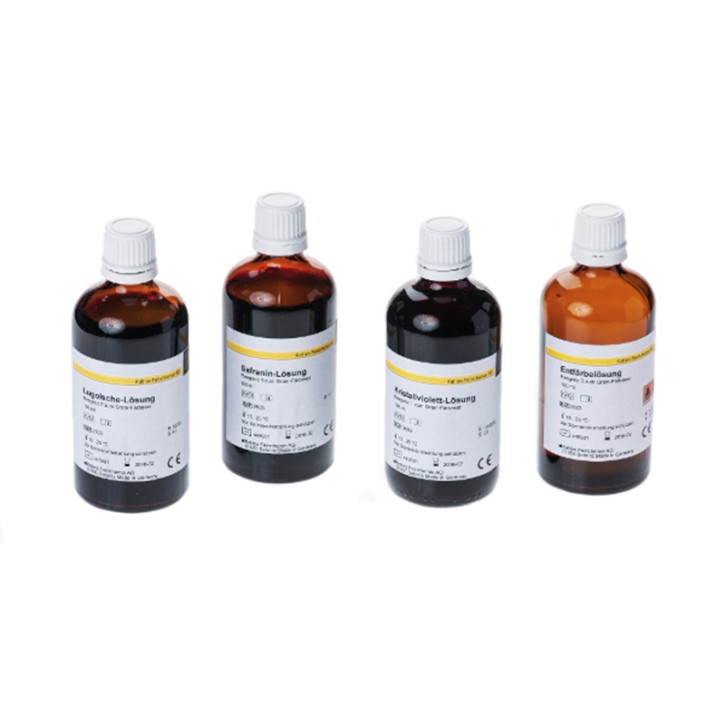
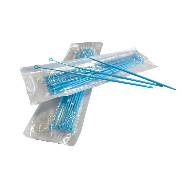
Background: Different bacteria have a different built of their cell membrane a circumstance which can be used to distinguish them by their staining color retaining properties. In general, bacterial cell membranes consist of the peptidal glycane mureine, a sugar polymere with multiple polysaccharide chains linked between each other . The single mureine polymeres are synthesized extraplasmatically, and form a shell of multiple layers aournd the bacteria called mureine sacculus. By the thickness of this mureine sacculus one can distinguish gram negative bacteria (not staining color retaining; few mureine polymere layers) and gram-positive staining bacteria (staining-color retaining; thick murine polymere layers). The technique of Gram staining was developed by the Danish biologists Hans Christian Gram (1853-1938) using a socalled differential staining with multiple steps (1) crystall violet und solution after Lugol, (2) wash out and fixation with ethanol, and (3) counter-staining with safranin-oxalate (red). Crystall violett binds to the acidic groups of the cytoplasm; Lugols staining contains iod forming intracellular not water-soluble complexes with the bound crystall violet. Ethanol (96%) drives those complexes back into solution and washes them out of the bacteria cells if not held back by the thick mureine polymer complexes of the Gram-positive bacteria. The counter-staining with safranine-oxalate colors both gram-positive as well as gram-negative bacteria, so that finally the former present with dark-violet staining, and the latter in red. The differentiation of gram-positive Vs. gram-negative bacteria is of significant evidence in the identification and differentiation of possibly pathogenic bacteria, as several antibiotics target the bacterial cell membrane synthesis (the mureine polymere synthesis) and in such case may be bacteristatic or bacteriozid ONLY against gram-positive species. Short protocoll: - Using an inocculation loop or a fine pipette tip, put one to two drops of an inocculate on a fatfree slide and generate a thin film using a clean cover slip. - Airdry at least 15 min - fixate gently with heat (coagulation of the protoplasmatic proteins fixates the cells on the slides surface - staining and washing steps 1 to 4 (1 min each..see IFU) -air-dry after sucking off water from the slide with filter paper -examine under the microscope

